Spine Brain Neurological and Neurosurgery Experts at Palm Springs Area
Chronic Sequestered Herniated Disk or Spinal Tumor?

60+ yo man who presented to the emergency room twice with approximately 5 days history of intractable pain, with burning sensation starting from his lower back with radiation to both lower extremities and involving the thigh and calf with the pain worse in the left compared to right, keeping him up at nights with constant cramping of his legs and together with weakness and progressive difficulty with gait and balance unable to walk independently.
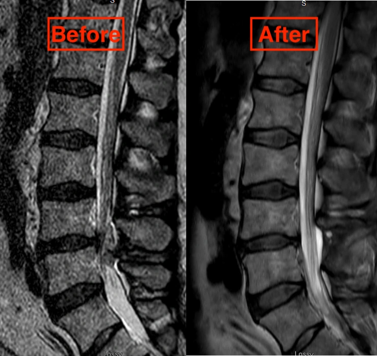
MRI scan of lumbar spine revealed a large contrast enhancing epidural mass at L4-L5 level, eccentric to the left resulting in severe stenosis and compression of thecal sac/spine and nerve roots.
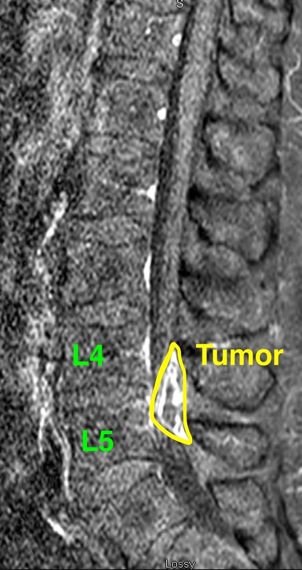
With diagnosis of Intradural-Extramedullary tumor he was taken to the operating room for surgical resection of this mass.
Tumor is well delineated here in contrast enhanced MRI of L spine.
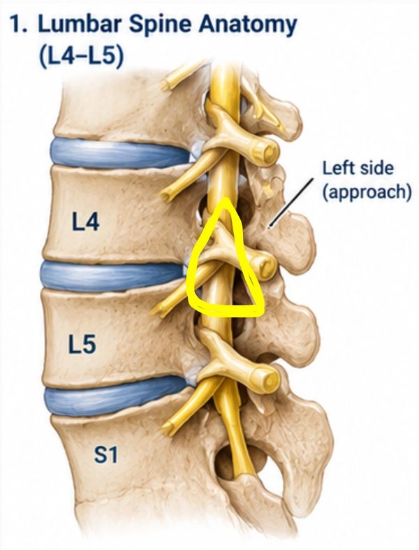
Surgical approach consists of left sided L4-L5 laminotomy to expose the tumor which is commonly performed for resection of a herniated disc where part of the lamina of L4 and L5 is removed with exposure of the herniated disc and its removal preserving the spine and nerve roots.
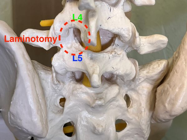
Here we use a spine model to orient you to the surgical field and view through the microscope.
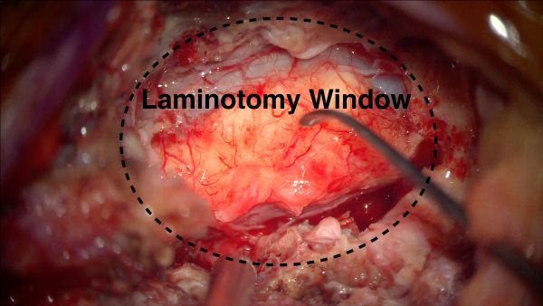
Laminotomy Window and you can see the tumor well on top of spine.
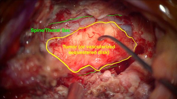
Laminotomy Window and you can see the tumor well on top of spine.
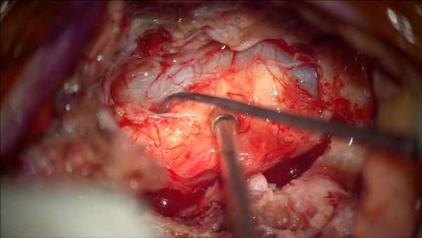
Tumor is dissected off the thecal sac of spine without dura tear or injury using micro-instruments (watch the video for technique)
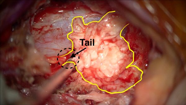
Once it is mobilized away from spine, it appeared that it arrises from L4-L5 disc with its tail incorporated with and arising from the L4-L5 annulus (watch the video for technique).
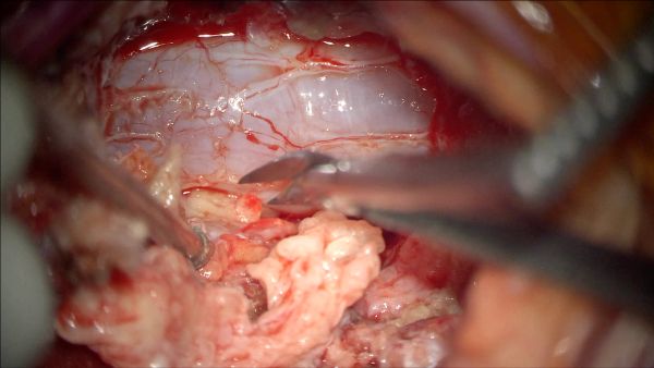
The mass was removed completely all the way to point of its probable extrusion from the annulus fibrosis of the L45 disc (watch the video for technique).
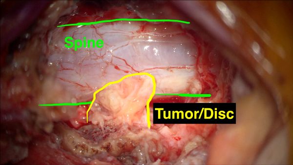
After removing the bulk of the mass, you can see its tail, or rather, where it had extruded from the L4-L5 disc space. This last portion is carefully removed without injury to the annulus fibrosis of the disk which could result in further disc herniation in the future(watch the video for technique).
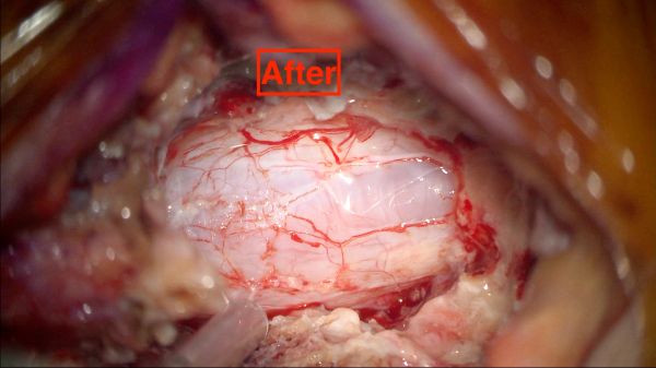
Tumor is completely removed off the spine. Although the official pathology of this mass was that of a fibrovascular tumor, it appears that it was a large herniated and sequestered disk. This sequestered or chronic herniated disc fragment had most likely undergone "organization" and neo-vascularization, which can look exactly like a fibrovascular mass or tumor.
Post Operation:
Post operative MRI Showed complete resection of this mass which was called a fibrovascular tumor (or in my judgment, a vascularized chronic sequestered herniated disc).
Patient did well post operatively with immediate resolution of pain, and gradual improvement of his muscle strength going from walking using a walker to complete normalization of his gait/balance/and strength, walking independently.
Watch the full video of the operation on Youtube link: https://www.youtube.com/watch?v=VzSS6poOx5I
Presentation by:
Look for the next monthly Case Presentation here.
Farhad M. Limonadi, MD